Detecting antigens and antibodies is a very important function of ELISA. The traditional method for detecting antigens or antibodies in biological fluids is known as ELISA. It is an extremely sensitive technique.
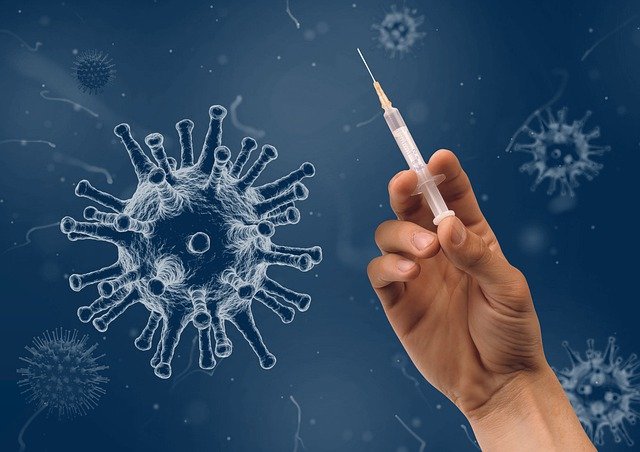
Image by Wilfried Pohnke from Pixabay
This means that even a few molecules of antigen or antibody can be detected, making it possible for very small quantities of substances to be detected. The sensitivity of ELISA is due to its specificity, accuracy, and reproducibility. ELISA also has a wide range of applications and can detect many different substances in different types of fluids.
In ELISA, antigens or antibodies are attached to a plastic or glass plate surface. This process is called coupling. After the antigen or antibody has been coupled to the surface, excess material is washed away. A solution containing an enzyme that recognizes and binds to the antigen or antibody is added to the plate. An antibody placed against the enzyme is required for this reaction to occur. If specific proteins are present in the samples, they will bind to their corresponding antigens on the plate surface, causing a visible reaction that the eye can see.
The ELISA technique can detect antigens or antibodies in different biological samples such as serum, plasma, urine, tissue culture, and cell culture supernatants. The ELISA assay has many advantages: it is more sensitive than other assays, gives fast results, does not require sophisticated equipment, does not damage the protein antigen, and can be easily automated for high throughput screening.
What are Antigens?
Antigens are foreign substances that stimulate an immune response. An antigen is any substance that can stimulate the production of antibodies or a T-cell response. Antigens can be proteins, carbohydrates, or glycoproteins. They are usually found on the surface of cells or viruses. Bacteria and cancer cells contain antigens on their surfaces that the immune system recognizes as foreign. This recognition causes our bodies to create antibodies that attack these foreign substances. Some antigens are proteins, and some are molecules that have been chemically attached to proteins.
These antigens fit into special areas of the lymphocytes called antigen receptors, which initiate an immune response when they bind to the antigen. The antigenic material is generally introduced by injection either under the skin or subcutaneously. The lymphatic system absorbs the injected material from where it is carried to the organs where immune responses are formed.
What are Antibodies?
Antibodies are molecules that the body produces in reaction to an antigen. Antigens are molecules that cause the immune system to respond. Antibodies will only bind to antigens of the same type if they have the same binding sites. The binding sites are also called epitopes. Antibody binding sites are short chains of amino acids, and they can be as small as four amino acids or as large as 20 amino acids. The vast majority of antibodies in the human body are Y-shaped, but they can also be straight or L-shaped or even branched and multi-armed.
Antibodies can neutralize pathogens and toxins by binding them tightly and blocking their activity. This is called opsonization. Once a pathogen or toxin has been opsonized, it can be more easily swept up by phagocytes (a type of white blood cell) and destroyed. Antibodies can also activate other parts of the immune system, such as complement proteins, to destroy pathogens or toxins directly. Antibodies can bind to receptors on cells, activating them against their usual function. This is why lots of people are opting for therapies that remove harmful and corrupted antibodies from the body. A popular way to do this is plasmapheresis treatment which includes a process of removing plasma from blood and then filtering it to remove antibodies. After this, the purified plasma is returned to the body.
How Does ELISA Detect Antibodies and Antigens
ELISA is a very sensitive test that is so accurate that it allows the detection of one antibody or antigen in a solution containing millions or billions of the substances to which they are directed. The test works by attaching an antigen onto a plate and allowing the sample to flow through the plate. If antibodies are in the sample, they will bind to their antigen on the plate. The antibodies then become visible when using a secondary antibody that causes them to glow when exposed to light.
There are several ELISA antibodies available for detecting antigens and antibodies. We would be looking at these IDS antibodies, Enos antibody, and FAP antibody.
IDS Antibody
The IDS antibody is also known as Anti-Iduronate 2 sulfatase. The antibody reacts with human samples and has Rabbit as its host. The IDS antibody contains each of its vial containing 5mg BSA, 0.9mg NaCl, 0.2mg Na2HPO4, 0.05mg Thimerosal, 0.05mg NaN3, and has no cross-reactivity with other proteins.
Enos Antibody
Enos Antibody has a size of 100μg/vial with clonality that is Polyclonal. Each vial contains 4mg Trehalose, 0.9mg NaCl, 0.2mg Na2HPO4, 0.05mg NaN3 and has Rabbit as its host. The Enos antibody has No cross-reactivity with other proteins.
Western blot analysis of NOS3 using an anti-NOS3 antibody (A01604-2) (https://www.bosterbio.com/anti-enos-nos3-picoband-trade-antibody-a01604-2-boster.html)
FAP antibody
FAP anybody has a size of 100μg/vial and has Rabbit as its host. FAP antibody reacts with mice and rats. Each vial contains 4mg Trehalose, 0.9mg NaCl, 0.2mg Na2HPO4, 0.05mg NaN3. FAP antibody is Polyclonal in clonality and has no cross-reactivity with other proteins.
Western blot analysis of FAP using an anti-FAP antibody (A00422-1)(https://www.bosterbio.com/anti-fap-picoband-trade-antibody-a00422-1-boster.html)
Related Posts: